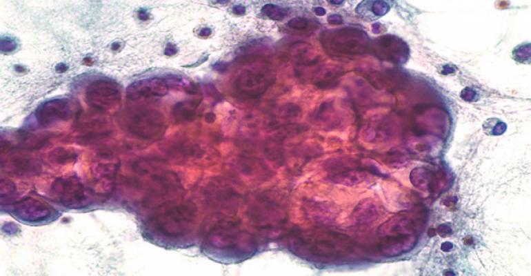
Pleural effusion is an abnormal accumulation of fluid present in the pleural cavity due to an imbalance mechanism of excessive fluid production or decreased lymphatic absorption or both. Normally the amount of pleural fluid is around at 0.1 ml/kg to 0.3 ml/kg and acts as a minimal lubricant for both pleural surfaces. As the most common pleural disease, it causes high mortality and morbidity and affects more than 1.5 million patients per year in the U.S. In Indonesia, 3,000 pleural effusions were diagnosed within January 2017 until February 2019, according to the medical record in Cipto Mangunkusumo National Referral Hospital.
Pleural effusion occurs as a result of the varied underlying health problems primarily involving the pulmonary or systemic system, which may affect the clinical symptoms, and further examination based on its aetiologies. Inflammation in pleural effusion often causes localised and severe pain with breathing or a cough; patient with systemic obstruction caused by congestive heart failure complains asymptomatic or breathlessness associated with its fluid volume. The build-up of fluid in pleural effusion can be caused by several mechanisms including increased pleural membrane permeability, increased pulmonary capillary pressure, decreased negative pressure in the pleural cavity, decreased oncotic pressure, and obstruction of the lymphatic vessels.
Classification of pleural effusion
Pleural effusion can be classified by the modified Light’s criteria. If the ratio of pleural fluid protein to serum protein is more than 0.5; the ratio of pleural fluid lactate dehydrogenase to serum LDH is more than 0.6; and/or the pleural fluid LDH level is more than 2/3 the upper limit of the normal level of serum LDH; at least one of the criteria is met and it is considered as exudative effusion. This condition can occur due to infections and inflammatory disorder such as tuberculosis, pneumonia; malignancy; hemothorax; parapneumonic effusion; and lymphatic obstruction. However, transudates are mostly caused by systematic conditions like congestive heart failure, nephrotic syndrome, liver cirrhosis, hypoalbuminemia; which alter the hydrostatic or oncotic pressure in the pleural cavity. Other less common causes are drug-induced, pulmonary embolism, oesophageal rupture, and post-radiotherapy.
Comprehensive diagnostic approach
Some patients presenting a pleural effusion can be asymptomatic or complain about dyspnoea, cough, and/or pleuritic chest pain; a thorough medical history may help to differentiate aetiologies from cardiovascular or other causes such a history of cancer, recent trauma, chronic hepatitis or an occupational history, and their detailed recent medication. A precise physical examination should be performed to determine if the clinical sign suggests an effusion like dullness to percussion.
Meniscus sign of pleural effusion can be detected by chest radiographs in the presence of 200 mL of fluid on posteroanterior radiography and in any case 50 mL of fluid is required on lateral radiography. Computed tomography (CT) can be used to evaluate effusions, which are not apparent in plain radiography, differentiating between the pleural fluid and pleural thickening, and provide clues to select the empyema’s site of drainage. Loculated or small amount pleural effusion, even in detecting pleural effusion in the critical care setting can be detected accurately by Ultrasonography. Its guidance is more sensitive in detecting pleural fluid septations, useful for established diagnosis and for marking a site of thoracentesis.
Thoracentesis is a minimally invasive procedure indicated to all unilateral effusion of unknown origin for diagnosis and relieving symptoms in patient. The effusion sample from this procedure can be analysed to differentiate its mechanisms as transudative or exudative effusions by using modified Light’s criteria.Pleural fluid needed to analyse is approximately 20-40 ml; then it will be extracted within four hours. A reddish pleural fluid appearance shows the presence of the blood like in trauma, pulmonary embolism, or malignant disease; if it comes after a prolonged period it will alter with a brownish tinge appearance. The presence of malignant effusion, tuberculous pleuritis, oesophageal rupture, or lupus pleuritis can be shown by a low glucose amount (30-50mL) and mostly with a low pleural pH level (less than 7.2). In most cases, higher LDH will present in exudative effusion as ongoing inflammation, whereas the decreasing LDH concentration showed in a cessation of the process in inflammation.
The predominance of white cells in differential cell count gives guidance to establish aetiologies; neutrophils caused by pneumonia or empyema, eosinophilia presents in pneumothorax, haemothorax, or asbestosis, mononuclear cell as the sign of chronic inflammatory, then lymphocytes will present in cancer and TB infection. Gram stain and culture in pleural fluid useful for detecting chronic illness or suspect TB. Since Indonesia has become a high burden country for tuberculosis, other promising diagnostic tools may be proposed as a proper index for TB diagnosis by the use of adenosine deaminase (ADA) that are more reliable, easy, rapid, and cost-effective.
Computed tomography (CT) can be used to evaluate effusions, which are not apparent in plain radiography, differentiating between the pleural fluid and pleural thickening, and provide clues to select the empyema’s site of drainage.
Diagnostic value of Adenosine Deaminase (ADA) in tuberculosis body fluid
The deamination of adenosine to inosine and of deoxyadenosine to deoxyinosine has been catalysed by ADA enzyme as a marker of cell-mediated immunity. ADA will elevate in empyema, whereas ADA was found to be high in M. tuberculosis infection. However, there is no significant difference in the use of total ADA and isoform ADA2 in clinical practice.
ADA analysis can be performed in the presence of exudate in pleural, ascitic and CSF with increased cell count of lymphocytosis using the Giusti method. Approximately 0.02 ml sample of body fluid and 0.2 ml of an adenosine solution were placed into a test tube, subsequently incubated at 37ºC for 60 minutes. Then, a phenol-nitroprusside and a hypochlorite solution will be added and incubated at 37 degree Celsius for 15 minutes. A Spectrophotometer at a wavelength of 620 nm will be used to read the amount of ammonia liberated by ADA action by converted to U/L.
ADA assay for detecting tuberculous pleurisy
The usefulness of ADA in pleural fluid for diagnosing tuberculous pleurisy had been investigated in some studies. Qureshi et. al (2018) reported 330 (93.75 per cent) out 352 TB pleurisy cases showed ADA levels 40 IU/L and above with a sensitivity 93.75 per cent, specificity 91.42 and positive predictive value 98.21 per cent in an area of high prevalence for tuberculosis, whereas in Auckland, New Zealand, as a low incidence setting, the median pfADA in 57 TB pleural effusion remains significantly higher (p<0.001) than in 1580 non-TB pleural effusion, respectively 58.1 U/L and 11.4 U/L presented by Blakiston et. al (2018).
The most widely used cut-off for tuberculous pleurisy diagnosis is approximately 40 IU / L. Study by Yusti G et al (2018) reported HIV patients with diagnosis of TB showed a median value of ADA of 70 IU/L (interquartile range (IQR) 41-89) and the non-TB group a median of 27.5 IU/L (IQR 13.5-52), hence ADA level determination could be useful to diagnose pleural tuberculosis in HIV infected patients.
Measurement of LDH/ADA ratio may be helpful for clinicians in distinguishing between tuberculosis with parapneumonic pleural effusion. Based on study by Wang J et. al (2017), the median pleural fluid LDH and ADA levels and LDH/ADA ratios in the tuberculosis and parapneumonic groups were: 364.5 U/L vs 4037 U/L (P < .001), 33.5 U/L vs 43.3 U/L (P = .249), and 10.88 vs 66.91 (P < .0001) respectively, showed the pleural fluid LDH/ADA ratio in the tuberculosis group was significantly lower than in the parapneumonic pleural effusion group.Prior studies have noted the importance of ADA as an useful diagnostic tool for the management of tuberculous pleurisy as an exudative pleural effusion.
Conclusion
Pleural fluid adenosine deaminase (ADA) can be a reference guidance to diagnose tuberculous pleurisy according to significant increasing level that has been found in tuberculosis infection as an exudative pleural effusion.
References available on request.

